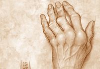
Rheumatoid Arthritis,
Hands
In the
hands, the metacarpophalangeal (MCP), proximal interphalangeal
(PIP), and thumb interphalangeal (IP) joints are involved
most frequently. The distal IP (DIP) joints are involved
only in the presence of coexisting MCP or PIP disease.
Tenosynovitis of the flexor tendons causes a reduction
in finger flexion and grip strength. Nodular thickening
in the tendon sheath also may produce a trigger finger.
With
progression of arthritis, characteristic deformities
of RA become apparent. These include ulnar deviation
of the fingers at the MCP joints, subluxation of the
MCP joints with the proximal phalanx slipping to the
volar side of the metacarpal heads, hyperextension of
the PIP joint with flexion of the DIP joint (swan-neck
deformity), flexion of the PIP joint with hyperextension
of the DIP joint (boutonni?re/button-hole deformity),
Z-shaped deformity of the thumb from subluxation of
the first MCP joint and compensatory hyperextension
of the IP joint, and drooping of the ring and little
fingers resulting from rupture of the extensor tendons
at the point of crossing the inflamed eroded ulnar styloid.
In
the wrist, early stages of RA cause tenosynovitis of
the extensor tendons, forming a swelling over the distal
wrist. The ulnar styloid may become tender, which indicates
inflammatory synovitis. The distal end of the ulna tends
to sublux dorsally, and the carpal bones sublux anteriorly
to the distal radius and ulna. Bony erosions and ankylosis
of the carpal bones also are seen and appear to be prominent
features in Asians.
Arthritis
typically has an insidious onset, with symmetric polyarticular
involvement of the small joints in the hands and feet.
Symptoms of pain and stiffness usually are present.
The classic persistent aching pain tends to have a diurnal
variation, ie, it is worse in the morning and eases
with activity. Stiffness also is more common in the
early morning after a period of inactivity. Stiffness
lasting more than 1 hour is fairly specific for inflammatory
joint disease.
Clinical
signs include joint swelling, muscle wasting, instability,
malalignment, and restriction of range of motion. Joint
swelling may be real or apparent, with real swelling
resulting from synovial thickening and joint effusion
in active synovitis and apparent swelling resulting
from malalignment.
In
addition, RA is a systemic disease and a number of important
extraarticular manifestations have been identified.
Fatigue, malaise, and weight loss are prominent features
and may reflect disease activity. Generalized osteoporosis
involving both the appendicular and axial skeleton is
common. A mild normochromic normocytic anemia commonly
is present, similar to anemia of chronic disease. However,
a degree of anemia lower than 10 g/dL is unusual. Felty
syndrome is the combination of neutropenia and splenomegaly
in RA.
Rheumatoid
nodules are small, firm, nontender subcutaneous nodules,
most often found over the proximal one third of the
ulna and at the olecranon. Nodules also may occur at
the fingers and thumbs (particularly in the dominant
hand) and elsewhere in the body. Nodules are strongly
associated with a positive rheumatoid factor.
Pleural
effusion and pleuritis are the most common pulmonary
manifestations. Pulmonary rheumatoid nodules are associated
with the presence of skin rheumatoid nodules and usually
are peripheral. They may cavitate but rarely calcify.
Multiple nodules on a background of pneumoconiosis are
known as Caplan syndrome. In addition, incidence of
pulmonary fibrosis and bronchiectasis is increased in
RA. Cardiac features include pericarditis and rheumatoid
nodules in the heart.
RA vasculitis frequently manifests as
obliterative endarteritis, with proliferation of the
intima in digital vessels resulting in nailfold and
digital infarcts. Several nerve entrapment syndromes,
such as the median nerve in carpal tunnel syndrome,
ulnar nerve in compression within Guyon canal, and posterior
tibial nerve in the tarsal tunnel, are more common in
RA. The eyes may show keratoconjunctivitis sicca and/or
scleritis. Sj?gren syndrome may occur together with
keratoconjunctivitis sicca.
Rheumatoid
factor is an immunoglobulin M antibody that is present
in 60- 80% of patients with RA at some stage during
the disease. However, rheumatoid factor is not specific
for RA and also is present in other connective tissue
diseases, infection, and autoimmune disorders. In addition,
rheumatoid factor is present in 1- 5% of people without
RA. Seropositive results are associated with nodules,
vasculitis.
Click
here to Read More about Rheumatoid Arthrits and Hands
Article
Sourced from eMedicine.com
|